
Hip: Illuminating Iliopsoas
"I have a tight psoas. I need to stretch the front of my hips."
I am sure we've all heard this plenty of times before from our clients but, when I hear these phrases my first reactions are:
- What do you mean by 'psoas'?
- Why do you hips feel so tight and when do you feel this tightness?
- Do you really need to stretch them?
- Are they being overloaded during postures or your exercise routine?
- Is there a way to improve your movement pattern to reduce this overload?
What intrigues me when clients present with this problem is why psoas gets so much attention and why other anterior hip muscles don't? You never hear a patient/client say "I have a tight sartorius or my pectineus is painful." Why does poor psoas get all the blame? It seems to be the only muscle ever addressed and I don't think that it's the only structure at fault. What I'd prefer to call it is anterior hip tightness/pain/dysfunction as I feel the hip needs to be address as an entire functional unit, not a single muscle.
Previously I've written blogs exploring the topics of:
- reducing TFL overactivity,
- identifying which exercises promote gluteal activity without overactivating TFL, and
- being specific about functional gluteal exercises.
The aim for this blog is around strategies for managing anterior hip and thigh tightness/pain. There are two avenues for this approach:
- Address the underlying dysfunction that caused the hip problem i.e. poor lateral stability, poor gluteal function, poor centering of the femoral head etc.
- Target the anterior hip to reduce pain and tightness.
When I started researching this blog I was hoping to illuminate iliopsoas and share wonderful knew information about this muscle.... but as I read and thought and cultivated ideas, I realised that it really shouldn't be all about iliopsoas. It should be about the entire anterior hip complex. Every hip complaint warrants a full hip assessment, not a quick fix approach. To that point I still wish to review the clinical anatomy of the anterior hip and with the help of Myotherapist and friend Kate Walters, discuss some treatment strategies for a "tight psoas".
Clinical anatomy

Visualising the muscles around the hip. IC - iliocapsularis, PA - psoas major, PI - psoas minor, R - rectus femoris, I - iliacus, S - Sartorius, PE - pectineus, TF - tensor fascia latae (Babst et al., 2011, p. 1733)
As you can see from the image above, there are many more structures to consider in our assessment of the anterior hip. Outside the joint, capsule, ligaments and labrum we have many muscles acting along different lines crossing the hip, originating from or attaching to and around the hip region. Just to name a few...
- In the anterior compartment there is sartorius, rectus femoris, iliacus, psoas major, psoas minor, iliocapsularis and pectineus.
- Laterally we also can see gluteus medius, glutues minimus and TFL.
- Also you can see the close proximity of the femoral nerve, artery and vein to pectineus, psoas major, psoas minor and iliacus.
When I look at an image like this I am reminded by how interconnected each of these structures are and that to claim one particular muscle is at fault in isolation, without assessing the others, will limit our clinical reasoning.
The muscles considered to act as deep stabilisers of the hip are quadratus femoris, gluteus minimus, the gamelli, obturator internus and externus, iliocapsularis and deep fibres of iliopsoas (Retchford., 2013). Therefore, if you are going to assess stability of the hip, considering the function of each of these muscles is important to remember.
If iliopsoas is a hip stabiliser, lumbar stabiliser and hip flexor, then it's behaviour and function is going to be impacted by all the other muscles acting as hip stabilisers or hip flexors. Coming back to my questions above, about why the hip flexors may be a source of trouble, I immediate wonder if iliopsoas is truely to blame or simply the manifestation of other control and mobility issues.
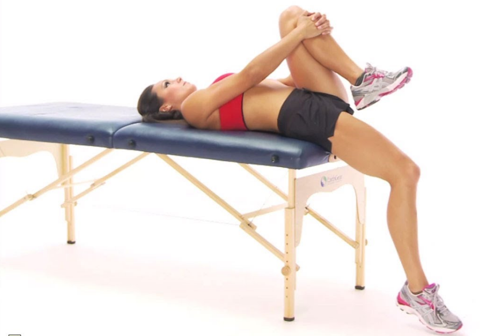
Image courtesy of Google Images
More than often I will find that patients with hip flexor tightness are not tight through this region, but using these muscles too much for stability during movement. Zach Long discusses how to assess if tightness is a true issue in his blog on hip flexor function. To assess someone's hip flexor demands a full hip assessment of structure, function and biomechanics. The main point I am trying to make here is that there is much more going on around the anterior hip that may contribute to sensations of "tightness" or "pain" that could be driven from structures other that iliopsoas.
Anterior hip assessment
- Functional:
- Control of movement and activation patterns during a stalk test looking at gluteal activation on the weight bearing leg and the movement of hip flexion on the non weight bearing leg.
- Assessment of their main aggravating movement.
- Palpation:
- Palpation of all the different muscles around the hip using movements such as hip rotation, hip adduction, hip abduction to confirm which muscles you are feeling.
- Palpation of the psoas muscles and iliacus around the abdominal region.
- Palpation of the bony landmarks - pubic symphysis, ASIS and along the line of inguinal triangle
- Muscle function:
- Squeeze test for adductor muscle function.
- Control of movement, pain provocation and stability during an ASLR test.
- Control of hip extension and pelvic rotation during a bridge or single leg bridge test.
- Range of motion testing:
- Prone hip passive extension.
- Thomas test position (iliopsoas) - Zach Long's blog (the Barbell Physio) covers the Thomas test technique in more depth as well as some exercise and manual therapy options.
- Ober's test (ITB and lateral thigh structures).
- Neurodynamics assessment:
- Prone knee bend (neurodynamic assessment of femoral nerve). Some studies have explored the correlation between reduced iliopsoas length (using the Thomas Test) and altered femoral nerve mobility (Prone knee bend test) and they found that: similarly to the interaction between neural mobility and muscular mobility in the upper limb or posterior thigh, that the Prone knee bend test and Thomas test are closely related (Anloague et al, 2015).
- SLR test (neurodynamic assessment of sciatic nerve) and the reason I say this is because if we cannot lengthen through the posterior chain, it will affect our ability to fold through the anterior chain and vice versa.
- Joint assessment:
- Flexion, abduction and external rotation (FABERS).
- Flexion, adduction and internal rotation (Quadrant).
- Compression and distraction.
- External and internal rotation in supine (90 degrees) and prone (more neutral hip position).
Clinical tips
So I began this blog by saying that iliopsoas often gets too much attention and blame. What I was trying to illustrate is the complexity of the anterior hip and that not all injuries are related to iliopsoas dysfunction. But there definitely are people who suffer from iliopsoas tightness and anterior hip pain, and if after you have completed your assessment, you feel these structures are at fault, here are some clinical tips.
When palpating this region I always start just above their ASIS or the crest of the anterior hip. The initial aim is to palpate the general area to check for tenderness through the abdomen and hip before going in deeper to palpate along the line of the iliopsoas.
My hand position of preference is with one hand over the other and fingers gliding down the fibre of the muscles. Depth is dependant on the patient's weight but it doesn't need to be forceful. Treatment of the iliopsoas is uncomfortable to a degree but this discomfort is often out-weighed by the response of releasing the iliopsoas muscle and surrounding region. Generally the iliopsoas muscles doesn't need a long treatment to gain relief, taking around 2-3 mins each side.
After treatment I recommend that the patient has the night to recover but depending on the severity of the injury or complaint, the patient should be able to get back into training or game day as soon as they feel comfortable (Kate).
treatment tips for iliopsoas
The most common position to begin treatment is in supine with legs straight. Try to palpate the abdomen with the pads of your fingers, not your fingers nails and using the patient's natural breathing rhythm to move deeper into the tissues is a great way to move through resistance.
Other positions you can try are with knees bent up and asking the patient to slide their leg into a straight position (hip flexion/extension) or to drop their knee to the side (hip external and internal rotation), while you maintain a constant pressure on psoas major. For a more progressed position, the patient can lie in the Thomas Test position with their foot supported on a stool or block, to have the hip on stretch during treatment. This position also gives great access to treatment of rectus femoris and other anterior thigh structures below the ASIS.
Once the muscle has been palpated the therapist will do techniques such as trigger point therapy, cross friction massage and stripping, or ask the patient to actively move through a direct (flexion/extension) or indirect (rotation) range (Kate).
Anterior hip Stretches
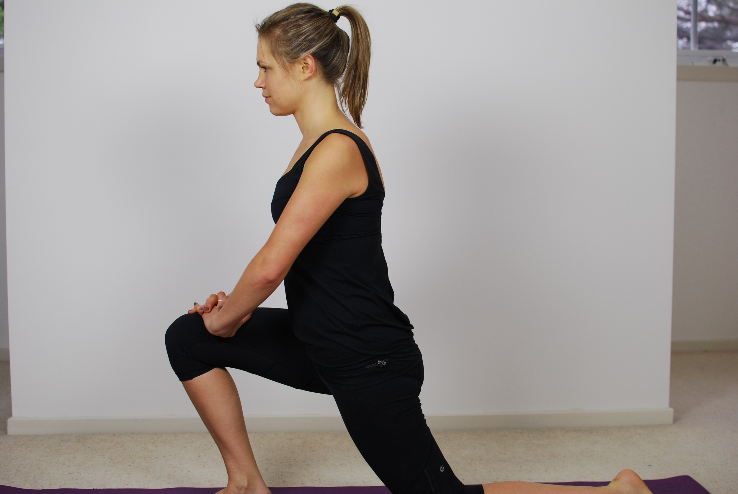
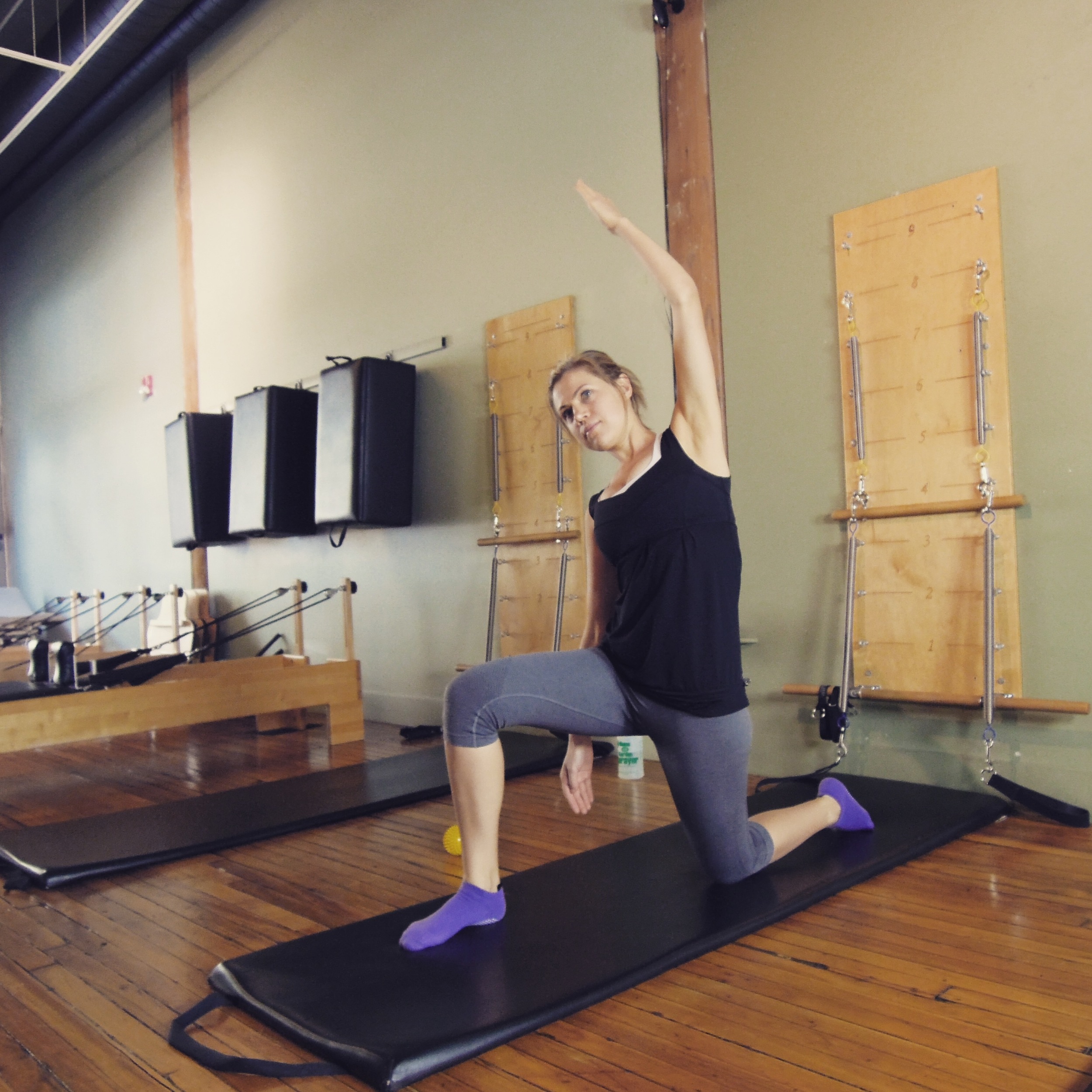
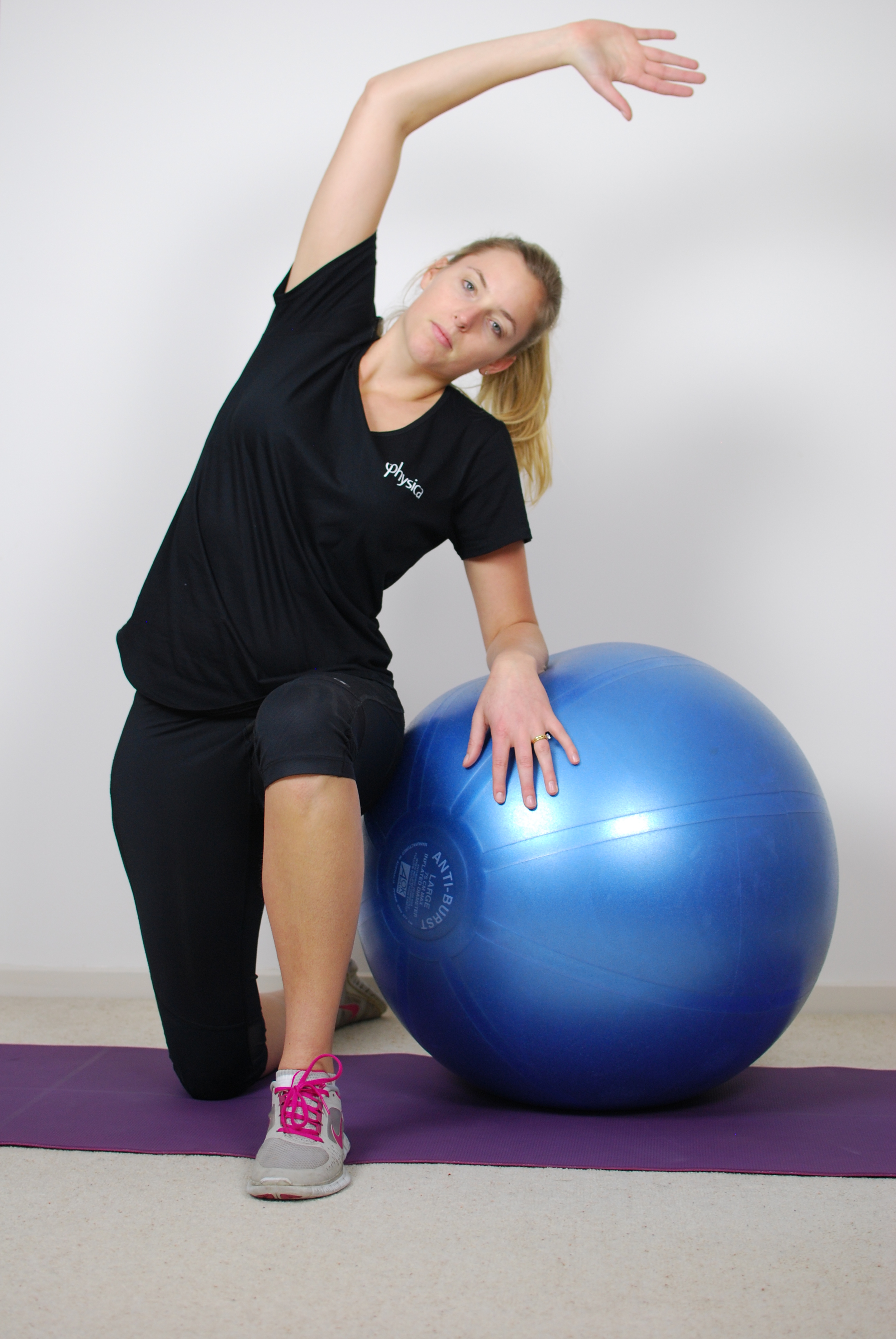
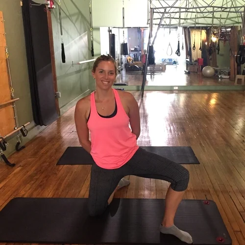
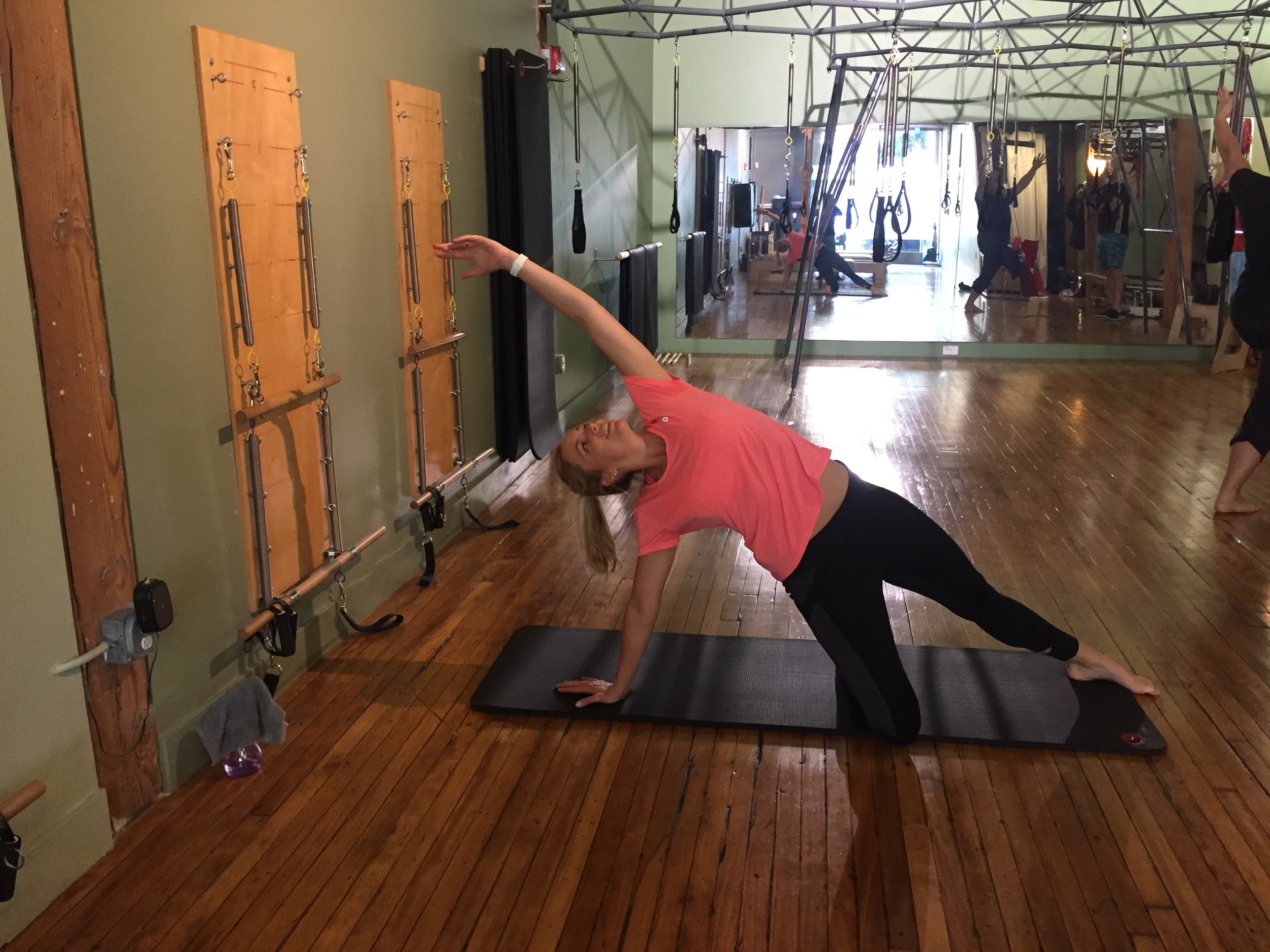

Image 1: your most common hip flexor stretch.
Image 2 & 3: adding an element of trunk lateral flexion into the stretch with or without support for balance. This is great if there is additional restriction through the lateral abdominal wall or lower back that can be contributing to loading the hip.
Image 4: half kneeling groin stretch (my favourite) is a stretch that targets the groin, anterior thigh and hip all in one go without forcing the stretching hip into too much extension. Some physios would say that if you have any labral issues anteriorly that stretching into end range extension might not place the best loads on the hip. This exercise doesn't knee much extension to feel great. Begin kneeling with knees apart and feet together then lift one leg up on a diagonal. Keep the pelvis posteriorly tilted and trunk facing forward and lunch sideways over the foot.
Image 5 & 6: some more unusual variations of and anterior hip stretch. The first is stretching the leg that is extended and I love this one again if you need to open the abdominal wall, lower back, and hip. The last picture is a strong quads stretch using the box on the reformer to hold the stretch and then move the hip into extension with the reformer. Similar to when people place their foot up against a wall behind them but you can move in and out of the stretch.
Anterior hip mobility exercises

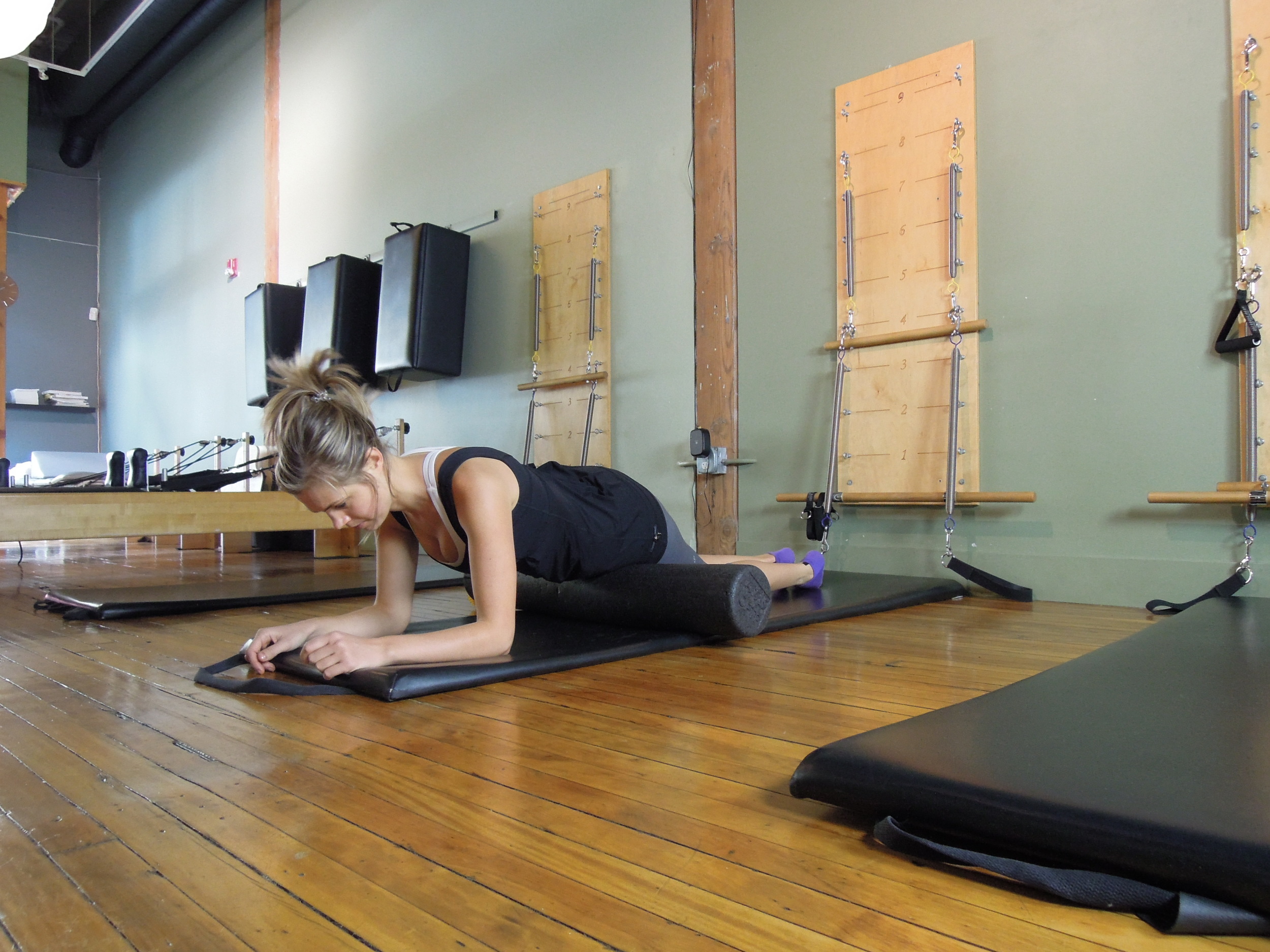
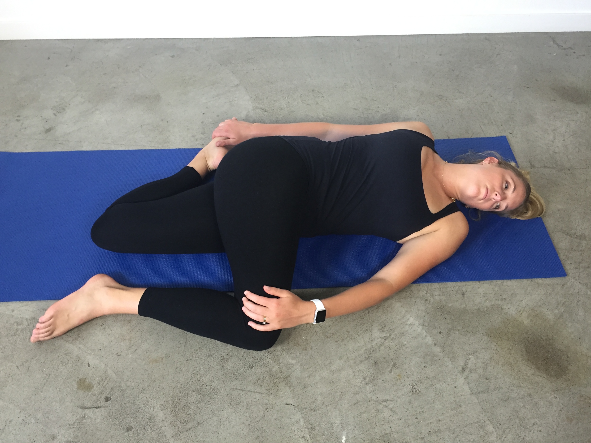
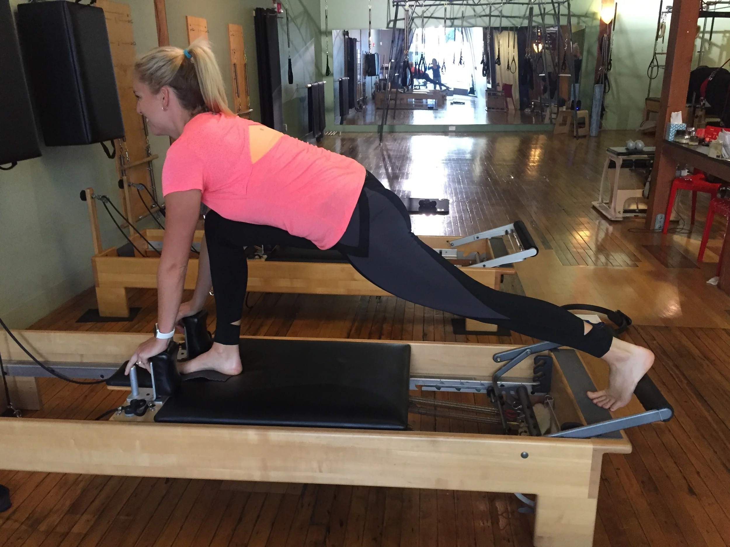
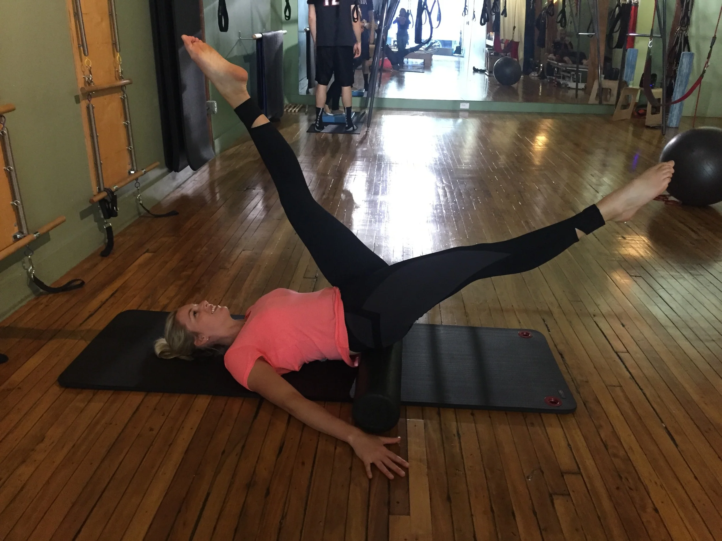
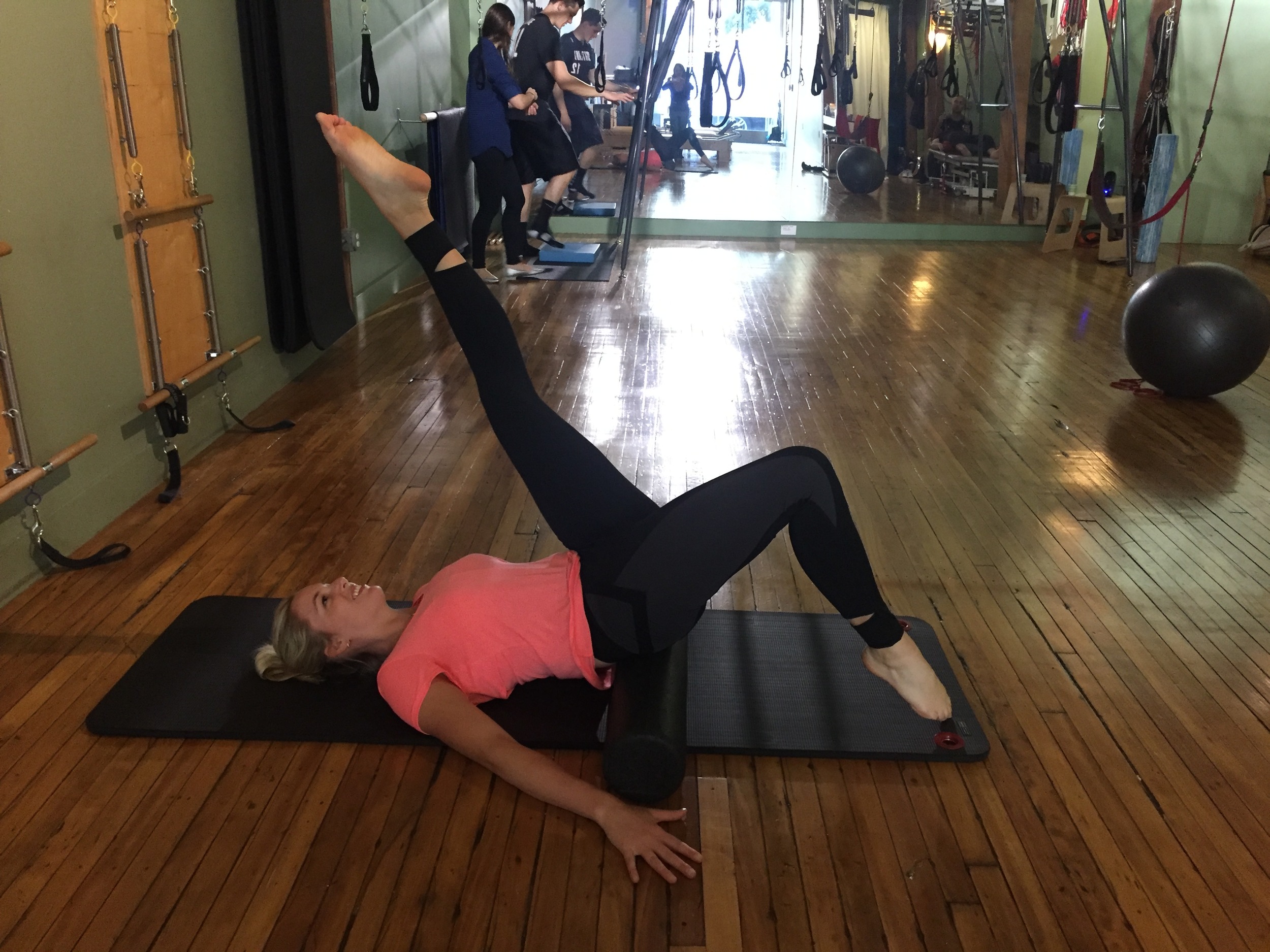
Image 1 & 2: using a spiky ball or foam roller to target the anterior hip. You can get into TFL, upper Rectus femoris and even Vastus lateralis.
Image 3: this stretch combines a quadricep stretch, lower back rotation stretch and upper thoracic rotation stretch (one the other side) all in one movement. What I love about this movement is that it shows people how connected the body is and that one segment may impact another e.g. lumbar rotation and gluteal mobility on my top leg affects the quads stretch on my bottom leg.
Image 4: Russian splits on the reformer - a great exercise for demonstrating hip hamstring mobility on one leg affects hip extension range on the other.
Image 5 & 6: simple hip control exercises with the pelvis supported on the foam roller. Focussing on scissors (pure hip flexion/extension) and bicycle kicks (bringing knee flexion and a quads stretch into the bottom of the movement).
My hope is that this blog has broadened your imagination about the many ways you can reduce the problem of anterior hip tightness and pain. There is a lot more to assess than the Thomas Test or treat than the kneeling hip flexor stretch. The aim for the second blog is focussing on enhancing abdominal muscle technique without loading the hips or lower back.
I want to thank Kate for her contribution to the blog. Kate has many many years of experience managing hip pain and sporting injuries and has brilliant soft tissue techniques. Its great to have a Myotherapist share on the blog as well. Here is a little bit about Kate.
Sian :)

Kate has been a Myotherapist for the past 8 years, practicing in the private setting and working with numerous elite teams, including Netball Victoria, Victorian Institute of Sports Diving, Melbourne Storm Rugby Club and St Kilda Football club. Kate is currently writing her degree of Health Science Complementary Medicine.
References:
Anloague, P. A., Somers-Chorny, W., Childs, K. E., Frankovich, M., Graham, C., & Birchfield, K. (2015). The Relationship between Femoral Nerve Tension and Hip Flexor Muscle Length. Journal of Novel Physiotherapies, 2015.
Babst, D., Steppacher, S. D., Ganz, R., Siebenrock, K. A., & Tannast, M. (2011). The iliocapsularis muscle: an important stabilizer in the dysplastic hip.Clinical Orthopaedics and Related Research®, 469(6), 1728-1734.
Karrasch, C., & Lynch, S. (2014). Practical approach to hip pain. Medical Clinics of North America, 98(4), 737-754.
Retchford, T. H., Crossley, K. M., Grimaldi, A., Kemp, J. L., & Cowan, S. M. (2013). Can local muscles augment stability in the hip? A narrative literature review. J Musculoskelet Neuronal Interact, 13(1), 1-12.